Revolutionary Ultrasound Device Allows for High-Quality Long-Term Imaging of Human Organs
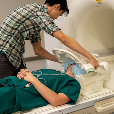
In the field of medicine, ultrasound is one of the safest methods for non-invasive examination of human organs. However, doctors have traditionally required technically complex equipment to send sound waves into a patient’s body and create images from the resulting echoes. Now, researchers at the Massachusetts Institute of Technology (MIT) have developed a compact ultrasound device that can be directly attached to the skin, allowing for continuous imaging of internal organs for up to 48 hours.
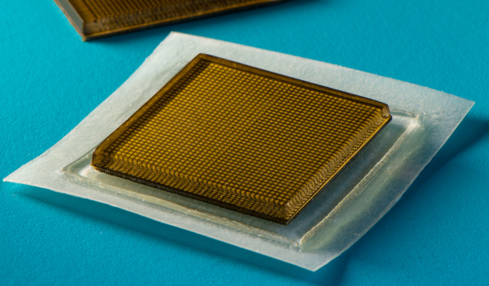
The device, which is about the size of a postage stamp, creates long-term images using ultrasound technology. The previous prototypes of the device had poor image quality due to the highly stretchable material used, which caused significant distortions. However, the current version has solved this problem with a new design featuring two layers. The sticky underside of the patch is highly flexible and conforms to the body, while the signal converter on top is relatively immobile, minimizing distortions.
In a practical test with multiple subjects, the researchers demonstrated the high image quality of the tiny ultrasound device. The subjects engaged in physical activity such as cycling and running, while the device continuously recorded high-resolution images of the most important blood vessels, heart, and lungs. Despite the high level of activity, the patch did not shift. Currently, the high-tech patch must be connected to an external device via cable for analysis of the recordings, but the next stage of development will allow for wireless transmission.
This breakthrough in ultrasound technology has the potential to revolutionize medical imaging, allowing for more accurate and continuous monitoring of internal organs. The device’s compact size and ease of use make it a promising tool for medical professionals in a variety of settings.